~ Shaan Gurnani's Organelles Project ~
~ Freshmen Biology - Mr. De - Period 6.8 ~
~ November 2007 ~
Cells and Organelles
Introduction:
Discovery of the Cell
- made possible by the microscope
- 1665 - Robert Hooke examined cork (named ‘little boxes’ cells)
- 1673 - Anton von Leewenhoek saw live cells
- 1838 - Matthias Schleiden said plants are made of cells
- 1838 - Theodor Schwann said animals are made of cells
- 1855 - Rudolf Virchow said cells come from other cells
Cell Theory
1.)All living things are composed of cells.
2.) Cells are basic unit of structure and function in an organism.
3.) Cells come from the reproduction of existing cells
A cell is very small, basic unit of life that is found in all functional organisms and is often called the building block of life. There are two main types: prokaryotic and eukaryotic.
Cell Diversity
- cells come in all shapes and sizes, specific to their function (> 200 different types)
Size:
- plant and animal cells, 10-50 micrometers
- bacterial cells, 0.2 micrometers
- as a cell grows, its volume increases faster than its surface area (think balloon) so…not enough surface (area) to take in nutrients or remove wastes
- cell will die if it could just keep enlarging – so it divides to make two more cells
Shape:
- shape of cell is important to its job in the body, i.e. long nerve cell, flat skin cells, globular white blood cell
- Prokaryotic: far more basic, no nucleus, DNA is in a ring, lacking other membrane bound organelles, usually found in bacteria
- Eukaryotic: more complex, have membrane bound nucleus to protect DNA, have membrane bound organelles (seperate parts with unique functions), usually found in plants and animals
A diagram showing the anatomy (all of the parts) of an animal cell:
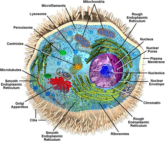
<-- picture from: http://micro.magnet.fsu.edu/cells/animals/images/animalcell.jpg
A diagram showing the anatomy (all of the parts) of a plant cell:

picture from http://www.ualr.edu/botany/plant3.gif
One of the Main/Major differences between an animal cell and a plant cell is that plant cells have a cell membrane and a cell wall while animal cells only have a cell membrane. Also, Plant cells also have chloroplasts a central vacuole. Animal cells have a flagellum, a lysosome, and a centriole.
An organelle is a specialized part of a cell. Each one has a specific function. All organelles work together as a unit to maintain cell life.
Outline and basic ideas:
- Extracellular fluid - fluid in external environment of cell
- Flagellum/Cilia (my choice) - propel cells through environment, move materials over cell surface
- Endomembrane system - group of several related organelles (following)
- Plasma membrane - cell membrane, protects cell from outside environment, selectively permeable
- Nuclear membrane - membrane of the nucleus, the "brain" of the cell
- endoplasmic reticulum (ER) - rough ER - prepares protein for export, smooth ER: makes steroids, regulates calcium levels, breaks down toxic substances
- Golgi Apparatus - processes and packages substances produced by the cell
- Vacuole - storage compartments for enzymes, waste products
- Cytoskeleton - network of long protein strands
- Mitochondria - energy "powerhouse"; transfer energy from organic compouds to ATP
- Ribosome - makes proteins
- Exocytosis/Endocytosis - import and export department of the cell
- Plastid (Chloroplast) - used by plants for photosynthesis
Extracellular fluid (ECF)
outside of cell fluid
- This is all body fluid outside of cells
- it is 1/3 of the body's liquid, 1/5 of body weight
- it is found in blood and in muscular and other body tissues
- within the body, this fluid, such as blood, is maintained at a certain pH (7.4) by buffers
- It is subdivided into three different parts:
- Interstitial Fluid - surrounds cells; 3/4 ECF
- Plama - part of blood; 1/4 ECF
- Transcellular fluid - part of digestive juices, mucus, and more

Flagellum/Cilium (my choice)
- plural: Flagella/Cilia
- make up a group of organelles called Undulipodium
- (notice that undulate is part of the group name)
- they are both very similar
- projections from the cell body
- are used for transportation (assist in movement)
- made up of microtubules
- Differences: (in function)
- Flagella:
- "whip-like" action to move the whole cell
- long and slender
- used to propel unicellular or small multicellular organism
- for example, allow sperm to move
- usually found in animal and protist cells, not plant cells
- can be more than one
- Cilia:
- waving action
- small hair-like and present in large numbers
- moves substances across the cell
- many
- may also be used as a sensory organ

Shown above: Escherichia coli bacterium with flagella
Picture from: http://www.monash.edu.au/news/assets/images/micro-robot.jpg
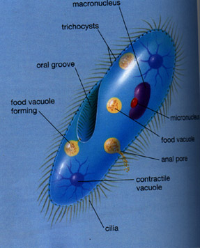
Picture from: http://sun.menloschool.org/~tbuxton/cells/f/animals/cilia_flagella/cilia.jpg
Endomembrane system:
- within eukaryotic cells
- this system is a collection of many membranes that create/seperate the various organelles
- they work together in the synthesis (production), storage and export of molecules
- the system includes the following organelles, which be covered in length following this:
- the plasma membrane -
- another term for the cell membrane -
- it is formed by a phospholipid bi-layer
- it seperates the cell from its environment
- regulates what can go in and out of the cell -
- (it is selectively permeable) -
- (this includes nutrients, waste, etc.)
- the nuclear membrane -
- also known as the nuclear envelope -
- is a membrane around the nucleus
- that encloses and protects genetic information
- Endoplasmic reticulum (ER) -
- synthesis (production) and transport organelle
- extension of the nuclear membrane
- Golgi apparatus
- packaging and delivery system for molecules
- vacuoles
-

The above image is::: a diagram that shows the various parts (organelles) of the endomembrane system in a eukaryotic cell.
Above picture is from::: http://upload.wikimedia.org/wikipedia/commons/thumb/0/00/Endomembrane_system_diagram.svg/612px-Endomembrane_system_diagram.svg.png
The Plasma Membrane:
- part of the endomembrane system
- also known as the cell membrane
- Structure:
- it is formed by a phospholipid bi-layer
- obviously, made up of several phopholipids
- As you can see in the diagram below,
- the hydrophobic fatty acids (the tails) are non-polar (water-fearing) and
- the hydrophillic head groups are polar (water loving - attracted to water)
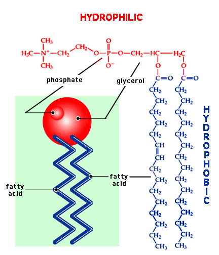
-
- This diagram shows the hydrophillic head and the hydrophobic tail of a phospholipid.
- The phospholipids form arrange in the following particular orientation because:
- there is H20 (polar) on the inside and H20 (polar) on the outside
- the fatty acids (tails) are hydrophobic and therefore face each away from the water and towards each other (the tails are attracted to each other from a phospholipid bi-layer)
- the head groups are hydrophillic and therefore face the water on the inside and the water on the outside
- Phospholipid bilayer is therefore a double layer of phospholipids
- a phopholipid are a type of lipids
 http://www.fz-juelich.de/inb/inb-1/datapool/page/28/Figure1-500.jpg
- ok so the following picture is a section of a phospholipid bilayer which forms the cell membrane...the blue things are proteins, they are membrane channels which allow things, such as water and nutrients,
- A unique feature about the cell membrane is that:
-
A cell membrane, unlike a cell wall (which is rigid), is fluid: it can move, it is flexible, and things can go through it
- This is all according to the Fluid Mosaic Model
-
- Fluid Mosaic Model
o lipid bilayer moves continuously and behaves like a liquid
o mosaic of proteins constantly changes as phospholipids move around and flip
- which was designed/discovered by S. J. Singer and Garth Nicolson
- the following image shows the cell membrane with all of its parts:
- (the phospholipid bilayer and several different types of proteins)
 http://en.wikipedia.org/wiki/Image:Cell_membrane_detailed_diagram.svg
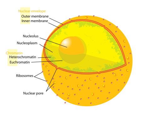
The Nuclear Membrane:
- The following image is a diagram of a nucleus in a human cell, from wikipedia.org: http://en.wikipedia.org/wiki/Image:Diagram_human_cell_nucleus.svg
- part of the endomembrane system
- the nuclear membrane -
- in eukaryotic cells
- also known as the nuclear envelope -
- is a double membrane (outer and inner)
- around the nucleus
- that encloses and protects genetic information
- it seperates what is within the nucleus from the cytoplasm
- there are several nuclear pores, marked on the diagram
- these are used to transport substances between the nucleus and the cytoplasm
- (allows materials to go into and out of the nucleus)
- Perinuclear space (space between the two membranes that composes the nuclear envelope)
- Outer membrane contains endoplasmic reticulum
- a nucleus is
- an organelle in eukaryotic cells
- the brain, or central control system, of the cell
- it contains all of the genetic information required (DNA)
Endoplasmic reticulum (ER):
 http://www.hhmi.org/askascientist/images/cell.gif
This image show location of the ER within the cell.
- Endoplasmic Reticulum (ER)
- part of the endomembrane system
- an organelle that is found in all eukaryotic cells
- network of many parts (tubes and vesicles)
- structure is similar to that of the plasma (cell) membrane
- intracellular highway, moves things through cell
- two parts/types
- covered in ribosomes (therefore it is rough)
- (the function of ribosomes is the production of proteins)
- synthesizes (produces) proteins
- transports proteins
- connected to the nuclear envelope
- Synthesizes steroids (thyroid gland)
- synthesizes lipids
- Regulation of calcium levels in muscle cells
- breakdown of toxic material by liver
- usage of carbs and {calcium] for energy
-

http://computer.act.ac.th/webproject5_2548/st/m53/division/Endoplasmic%20reticulum.jpg
Golgi Apparatus
- Part of the Endomembrane system
- in eukaryotic cells
- discovered by an Italian physician named Camillo Golgi in 1898
- basically it processes and packages,, materials (cell products such as macromolecules)
- and prepares for export out of cell through exocytosis
- (important for releasing materials from the cell)
- you can think of it as a mail center:
- preparing, processing, and packaging items
- to be sent out (of the cell, out of the post office) to another place
- Structure:
- six to seven flattened, membrane-bound sacs called cisternae
- located near the nucleus
The following image is a cell with the golgi apparatus label. I got the image from http://sun.menloschool.org/~cweaver/cells/c/golgi/cell.jpg

a closer look at strucutre, location, and parts of the golgi apparatus:: from http://micro.magnet.fsu.edu/cells/golgi/images/golgifigure2.jpg

Vacuole:
- last part of the endomembrane system
- eukaryotic cells
- found in most plant cells and in some animal cells
- are big in plant cells and small in animal cells
- sac bound by membrane
- small storage compartments in cell
- plays a role in releasing/exporting waste/not needed products out of the cell
- maintains turgor pressure which is important to keep plant cells rigidy and have them not wilt


First image (left) is a cartoon image: from::: http://www4.alief.isd.tenet.edu/cahowe/PreAPBio/Projects/Cell_files/slide0013_image053.jpg
Second image (right) is a microscopic image: from::: http://www.cellsalive.com/cells/vacuole.htm
Cytoskeleton:
- network of long protein strands located in the cytosol
- not surrounded by membranes
- made of microfilaments and microtubules
- Microfilaments
- protein threads of actin
- cell movement and muscle contraction
- Microtubules (conveyor belts around cell that move organelle around
- longest strands of cytoskeleton
- make up spindle fibers (role in mitosis and meiosis)t


I believe that in the left picture, the blue represents either the nucleus or the MTOC. Similarily, in the picture to the right, I believe that the purple sphere is the MTOC and the gold/yellow represents microtubules.
left picture from: http://content.answers.com/main/content/wp/en-commons/thumb/7/70/300px-FluorescentCells.jpg
right picture from: http://www.immediart.com/catalog/images/big_images/SPL_6_P780110-Fibroblast_cells_showing_cytoskeleton.jpg
Mitochondria:
- Mitochondria
o powerhouse of cell, uses ATP for energy, numerous in muscle cells and liver cells, have their own DNA
o two membranes
§ outer membrane is boundary
§ inner membrane is cristae, highly folded for increased surface area for reactions
picture from http://www.cartage.org.lb/en/themes/sciences/zoology/AnimalPhysiology/Anatomy/AnimalCellStructure/Mitochondria/mitochondria.jpg


Ribosomes
- do not have a membrane
- made/consists of both RNA and proteins parts
- made within nucleolus
- can be free floating or attached to rough ER (Endoplasmic Reticulum)
- make proteins, in a function called translation
- site of protein synthesis (free- within cell; RER (Rough Endoplasmic Reticulum) – exported out of cell)
Right image, ribosome protein from: http://publications.nigms.nih.gov/insidethecell/images/ch2_ribosome_proteinbig.jpg
Exocytosis/Endocytosis
 This animation shows exocytosis. http://academic.brooklyn.cuny.edu/biology/bio4fv/page/exocy.htm
Exocytosis:

http://www.linkpublishing.com/exocytosis5.jpg
The following picture shows both exocytosis and endocytosis:
endocytosis (import) is on the top and exocytosis (export) is on the bottom of the diagram
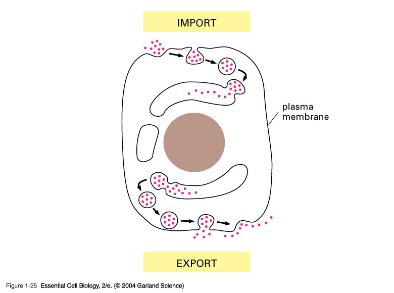
http://www.accessexcellence.org/RC/VL/GG/ecb/import_export.html
The import/export "department" of the cell:
Endocytosis:
- endo means "in"
- cells import materials (nutrients, etc.) from the external environment using Endocytosis
- as you can see in the above image, the cell captures the material in a vesicle (like a bubbles) and pinches it off from the cell (plasma) membrane
Exocytosis:
- opposite of endocytosis
- "exo" is similar to exit and means leave
- this is a process for exporting, or removing, substances such as waste and cell products from the cell
- a vesicle (bubble, holder) is created and it contains the substance
- it moves towards the membrane and eventually fuses with it
- the animation and the second image are both very good examples of exocytosis
Plastid (Chloroplast):
 <-- picture from http://biology.nuim.ie/graphics/chloropic_000.gif
basic model of the plastid
- found only in plant cells
- in charge of Photosynthesis, a process by which plants convert light energy into plant energy
- the reactants are CO2, H20 and energy from sunlight
- the products are glucose and oxygen
- also responsible for storage of starch
- surrounded by double membrane
- have their own DNA
- can store starch, fats, or pigments
- chloroplast convert solar energy to chemical energy
- contains thylakoids – flattened membranous sacs where photosynthesis occurs
- a granum is a stack of thylakoids
- stoma is the aqueous space
- Chlroplasts In an actual plant cell:
 http://en.wikipedia.org/wiki/Image:Chloroplasten.jpg
The two following images give a more in-depth look at the chloroplast, looking at various parts of it.
 http://www.daviddarling.info/images/chloroplast.jpg

http://www.ualr.edu/botany/chloroplast.jpg
Bibliography
- Campbell, Neil A., et al., eds. Biology Concepts & Connections. 5th ed. Pearson/Benjamin Cummings.
- Several websites
by Shaan Gurnani / period 6.8 / Mr. De


Comments (0)
You don't have permission to comment on this page.